BRUSATTE_ET_AL_Figure2

Diagram of brain case

Top row: Partial braincase of Timurlengia euotica in tree views (L to R: from the back, from below, and from the right side). Bottom row: Composite images of the brain case from CT scanning. Reconstructed brain in dark blue, inner ear in pink, nerves in yellow, and blood vessel in red. © Proceedings of the National Academy of Sciences
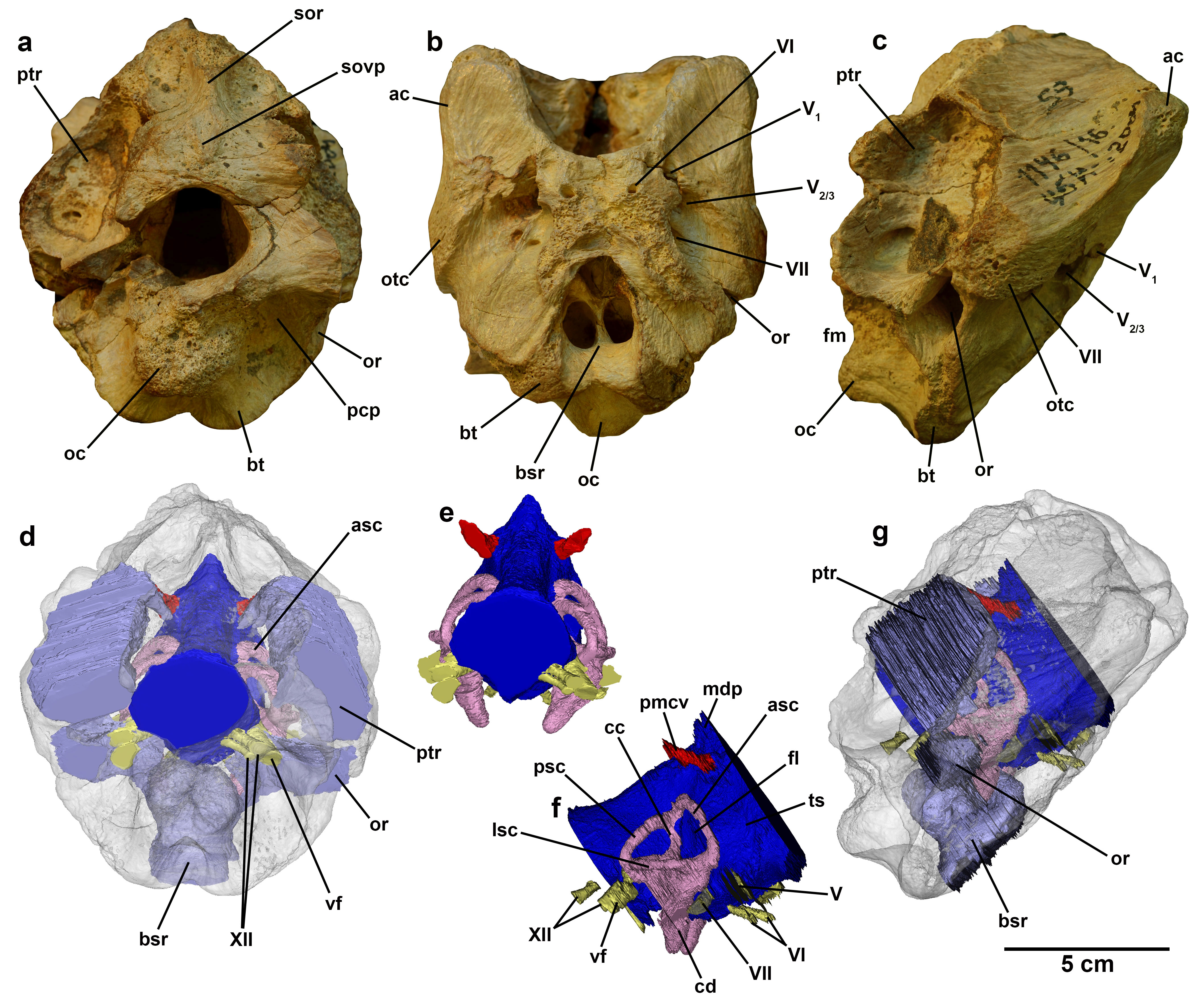
Top row: Partial braincase of Timurlengia euotica in tree views (L to R: from the back, from below, and from the right side). Bottom row: Composite images of the brain case from CT scanning. Reconstructed brain in dark blue, inner ear in pink, nerves in yellow, and blood vessel in red. © Proceedings of the National Academy of Sciences

What do you think?

Your email address will not be published.

You may use these HTML tags and attributes: <a href="" title=""> <abbr title=""> <acronym title=""> <b> <blockquote cite=""> <cite> <code> <del datetime=""> <em> <i> <q cite=""> <s> <strike> <strong>