November 8 is X-ray Day
Wilhelm Conrad Roentgen (1845 – 1923), a German physicist, discovered a new form of radiation at the University of Wurzburg in Germany, on November 8, 1895. While experimenting with cathode rays by passing an electric current through a glass vacuum tube covered with black paper, he noticed an unexpected green glow on a little screen covered with phosphorescent paint lying on his bench. He quickly realized that some mysterious invisible rays were leaving the tube, going through the black paper, and causing the screen to become luminous. These unknown, or “x” rays passed easily through wood, cloth and paper, and Roentgen showed that they could even pass through the skin and reveal the bones of the human hand.
Celebrate X-ray Day by donning your x-ray specs and take a closer look at the far-reaching implications of Roentgen’s Nobel-prize winning discovery.
Early x-ray tube
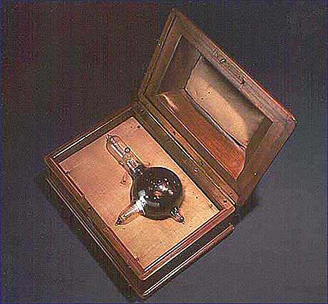
This tube was purchased from a private owner in Germany and presented to the Smithsonian Institution in 1956 by the General Electric Company’s X-ray Department of Milwaukee, Wisc. It is part of a very large and rich radiology collection at the National Museum of American History.
This is one of the first x-ray tubes used by Wilhelm Roentgen. The medical diagnostic and therapeutic implications of the x-ray were realized quickly. X-ray imaging remains the most widely used form of body imaging today.
Fluoroscope

In 1986, the National Museum of American History acquired this fluoroscope, one of perhaps only a handful extant, from a shoe store in northern Ohio. The mid-1930s vintage, walnut-cabinet machine was one of thousands produced by the Adrian X-Ray Company of Milwaukee, Wisc., a leading manufacturer of the devices.
The shoe-fitting fluoroscope used cutting-edge technology—the x-ray—to reveal the bones and soft tissue of the foot inside the shoe, ostensibly for a better fit. For three decades beginning in the mid-1920s, millions of children and adults in the United States, Europe and other parts of the world peered into the machines for an inside view of their usually wiggling toes.
Suited for Space

Image left by Mark Avino; right by Ron Cunningham, National Air and Space Museum
The history of this country is one of perilous voyages and uncharted territory, but few journeys have captured the imagination and hearts of the public as the race into space. The ingenuity and innovation that made space travel possible is celebrated in the exhibition Suited for Space. Rare and original photography, including unique x-ray images, reveal the modern technological marvel that is the spacesuit.
Chandra X-ray Observatory

An assortment of images from the Chandra Archive Collection: Preserving the Legacy of the X-ray Universe.
Fish Inside Out
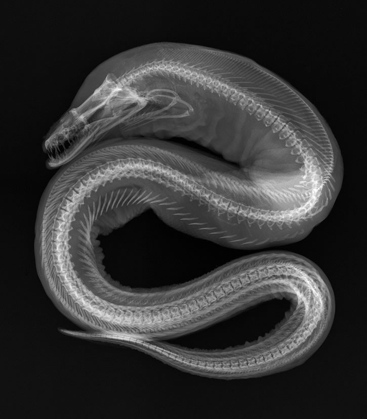
X-ray of of a Moray eel, Enchelynassa canina, courtesy National Museum of Natural History.
The Smithsonian’s National Collection of Fishes at the Natural History Museum is the largest and most diverse collection of its kind, with an estimated four million individual fish specimens representing more than 70 percent of the world’s fish species. The care, maintenance, and loan of these collections are the responsibility of a handful of museum specialists, including Sandra Raredon, who for 25 years has included radiology among her responsibilities. Her striking black-and-white radiographs, or x-rays, of fish, stingrays, eels, and seahorses—“anything with a backbone,” she notes—reveal the complex bone structure in a level of detail reminiscent of fine engraving and are the subject of the eerily beautiful exhibition X-ray Vision: Fish Inside Out.
X-ray diffraction

The gem pictured here is ametrine–part amethyst and part citrine. The most significant source of ametrine is from the Anahi Mine in eastern Bolivia. This pentagon-shaped ametrine weighs 91.93cts and was faceted by Jeff Gurecky. (Photo by Jeff Scovil)
Minerals are the basic geological building blocks of the Earth and solar system—studying the nature and behavior of minerals is essential for understanding the physical, chemical and biological processes that shape our planet. Researchers in the Department of Mineral Sciences at the National Museum of Natural History are examining the compositions and structures of minerals to understand a variety of processes, including how minerals control the distribution of chemicals and nutrients in the environment, the evolution of the Earth’s crust and mantle, and the causes of defects and impurities in the coloring of gems and minerals. X-ray diffraction is the definitive method for identifying minerals and other crystalline materials. X-rays are diffracted by the regular three-dimensional arrangements of atoms in crystals and a diffraction pattern can be used as a “fingerprint” for a crystal’s identification.
X-ray vision

From the American History Museum exhibition, “The Price of Freedom: Americans at War”
“We are all in it—all the way,” President Franklin D. Roosevelt told Americans during a radio broadcast on December 9, 1941. “Every single man, woman and child is a partner in the most tremendous undertaking of our American history.” Even for children, the reality of a nation at war could not be avoided. Many of their favorite characters from the funny pages and comic books went off to fight. However, that most American of heroes—Superman—never joined the Army. He was classified 4-F when his X-ray vision skewed the induction eye test, so he used his super powers to aid the war effort in other ways, such as encouraging kids to use their pennies for victory bonds and join the Junior Defense League.
Related posts:


Posted: 8 November 2013


